Inside Neuroscience: Imaging Your Unique Brain
Historically, functional brain imaging has assumed that our brains are all essentially built the same way, with cells performing similar jobs in basically the same positions. But new precision imaging techniques show that the brain is more like a fingerprint: people might have similar patterns and structures in the brain, but the fine details vary.
“Until very recently… we were just trying to treat ‘the brain,’ which is something that doesn't really exist,” said Nico Dosenbach, a professor of neurology at Washington University School of Medicine.

Nico Dosenbach
In a press conference titled “Precision Brain Mapping: A New Frontier in Personalized Neuroscience,” moderated by Dosenbach, researchers described methods for imaging the functional patterns of a single brain. These precision scans are revealing new, previously undiscovered connections and networks in the brain, some of which align with poorly understood medical conditions. These imaging techniques could one day help physicians personalize treatments for a range of psychiatric and neurological conditions.
Unique Structures Drive Executive Function
The brain’s prefrontal cortex is the home of executive function, the ability to control and coordinate many different networks and tasks occurring in the brain.

Zach Ladwig
“When we experience damage to the prefrontal cortex… we have trouble making plans, being able to update those plans flexibly when things go wrong, or being able to stick to those plans and not get distracted,” said Zach Ladwig, formerly a graduate student at Northwestern University and now a research scientist at University of Illinois Urbana-Champaign.
Traditional functional magnetic resonance imaging (fMRI) averages the data of many people together. While the technique reduces noise, it also erases features that are unique to individuals. To help see these unique features, Ladwig averaged many high-resolution scans of the same person. Comparisons between numerous individuals revealed similar but distinct patterns across the prefrontal cortex, where small patches corresponding to different networks were in slightly different locations. Averaging multiple people would make some patches disappear, and other patches appear much larger.
Precision fMRI can not only “allow us to find specific targets for each person, but also can establish new [therapeutic] targets,” said Ladwig.
Precision Imaging and Social Language
Rodrigo Braga
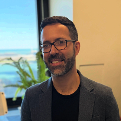
Rodrigo Braga, a neuroscientist at Northwestern University, is exploring similar questions in the inferior frontal cortex and its connections to the amygdala. The amygdala anchors a brain network called the “social cognitive network,” which fires when we think about what other people might be thinking or feeling.
By comparing high-resolution fMRI images of six individuals, Braga demonstrated that, similar to Ladwig’s work, each individual had language and social patches quite close together in the inferior frontal cortex, but the placement of these patches varied between individuals. In particular, he noted that one of these patches connected to the amygdala and the social cognitive network.
“We can now explain why is it that this particular part of the inferior frontal cortex has a social cognitive role, while the adjacent part is doing a different role, i.e., language,” said Braga. In other words, the connections to the amygdala matter more than the specific location on the brain when it comes to understanding the function of the inferior frontal cortex.
“Precision functional mapping is showing just how complex, how much more intricate the organization really is,” Braga said.
Newly Discovered Networks
Evan Gordon
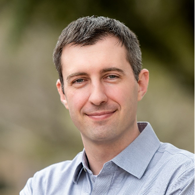
The uniqueness of the individual brain extends beyond just the mapping of known networks. Evan Gordon, a professor of radiology at Washington University School of Medicine in St. Louis, is using precision brain mapping to find previously unknown — and even individually unique — brain networks. He hopes that mapping an individual’s brain networks, and understanding if one or more networks are dysfunctional, could help treat neurological disorders.
“If you know which networks are doing what, which parts of the brain are doing what, and you want to modulate that function… now you have a target for that particular patient,” Gordon said.
Gordon previously published data revealing a novel network that he called the “SomatoCognitive Action Network,” or SCAN, which only became clear with precision fMRI. More recently, he’s looked deeper to see if he could find more unknown networks.
“Some of these new networks that we find appear to be only in a few people, or sometimes only in one person. These may reflect fundamental individual differences in how our brains work,” said Gordon.
Individualized Brain Networks in the Clinic
Gracie Grimsrud
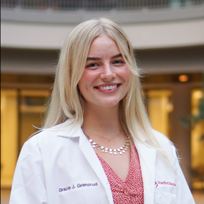
While brain injuries might be located at a specific region of the brain, many neurological and psychological conditions involve disruptions of brain networks. Gracie Grimsrud, a graduate student at Stanford University, is exploring the connection between Gordon’s recently discovered SCAN and how the drug Ritalin modulates the symptoms of attention deficit hyperactivity disorder (ADHD).
SCAN connects regions of the brain that handle bodily control, including movement and intention, as well as bodily functions like heart rate and pupil dilation. “I think of [SCAN] kind of as the brain's body orchestra conductor,” said Grimsrud, “because it's ensuring that both preparation for action and the action itself are all carried out in harmony.”
Many of the behaviors observed in people with ADHD, like impulsivity or difficulty initiating or maintaining a task, map onto the functions controlled by SCAN. So Grimsrud analyzed data from a study involving participants previously diagnosed with ADHD, and used high-resolution fMRI scans of their brains before and after taking Ritalin. In some participants, Ritalin helped control their impulsivity, while in others Ritalin did not help. In those patients responsive to the drug, imaging revealed a change in SCAN activity, while those who did not respond to the drug had no change in SCAN. In future studies, Grimsrud hopes to better understand how broader behavioral patterns coincide with imaging.
Targeting Therapy with Precision Imaging
Alik Widge, a clinical psychiatrist at the University of Minnesota, uses deep brain stimulation (DBS) to help patients with neurological conditions such as treatment-resistant depression. However, one of the biggest problems with DBS is figuring out exactly where to place the implants that carry electrical fields to the brain.

Alik Widge
In Parkinson’s disease, Widge noted, DBS has been quite successful in controlling tremors because monitoring the severity of tremors is an easy way to figure out if you’ve shaped the electrical field to affect the target brain circuits correctly.
But for conditions like depression or obsessive-compulsive disorder, it’s much harder to find that spot.
Widge believes that many of these neurological and psychiatric disorders are “disorders of stuck,” he said. “People are getting stuck in a behavior or thought loop.” And if that’s the case, then a type of test that observes “stuck-ness,” paired with precision imaging, might help physicians better tune DBS placement.
In a test, Widge gave patients who already had DBS neurostimulators a task that required behavioral flexibility. And he noted that when the stimulators were on, study participants were quicker — by just a few milliseconds — at switching tasks. Widge plans to test this technology to see if it helps with placing DBS implants in patients with severe anxiety or depression. The study has been derailed by NIH funding cuts, although the team still hopes to treat a few patients with philanthropic funding as a proof of concept.
While precision functional imaging is still a research technique, the presenters shared the view that the promise for the clinic is enormous. “Really, every brain's different, and we should treat your brain, not the brain,” said Dosenbach.