Brainy Zoom Backgrounds for Your Next Meeting
SfN collects scientific images from around the world from researchers who publish in our journals, JNeurosci and eNeuro. Both journals publish highly rigorous research representative of the breadth of neuroscience, and SfN members enjoy full access to the content of both journals. Explore the Zoom backgrounds below to discover images that have been published in JNeurosci and eNeuro.
SfN Zoom Backgrounds
-
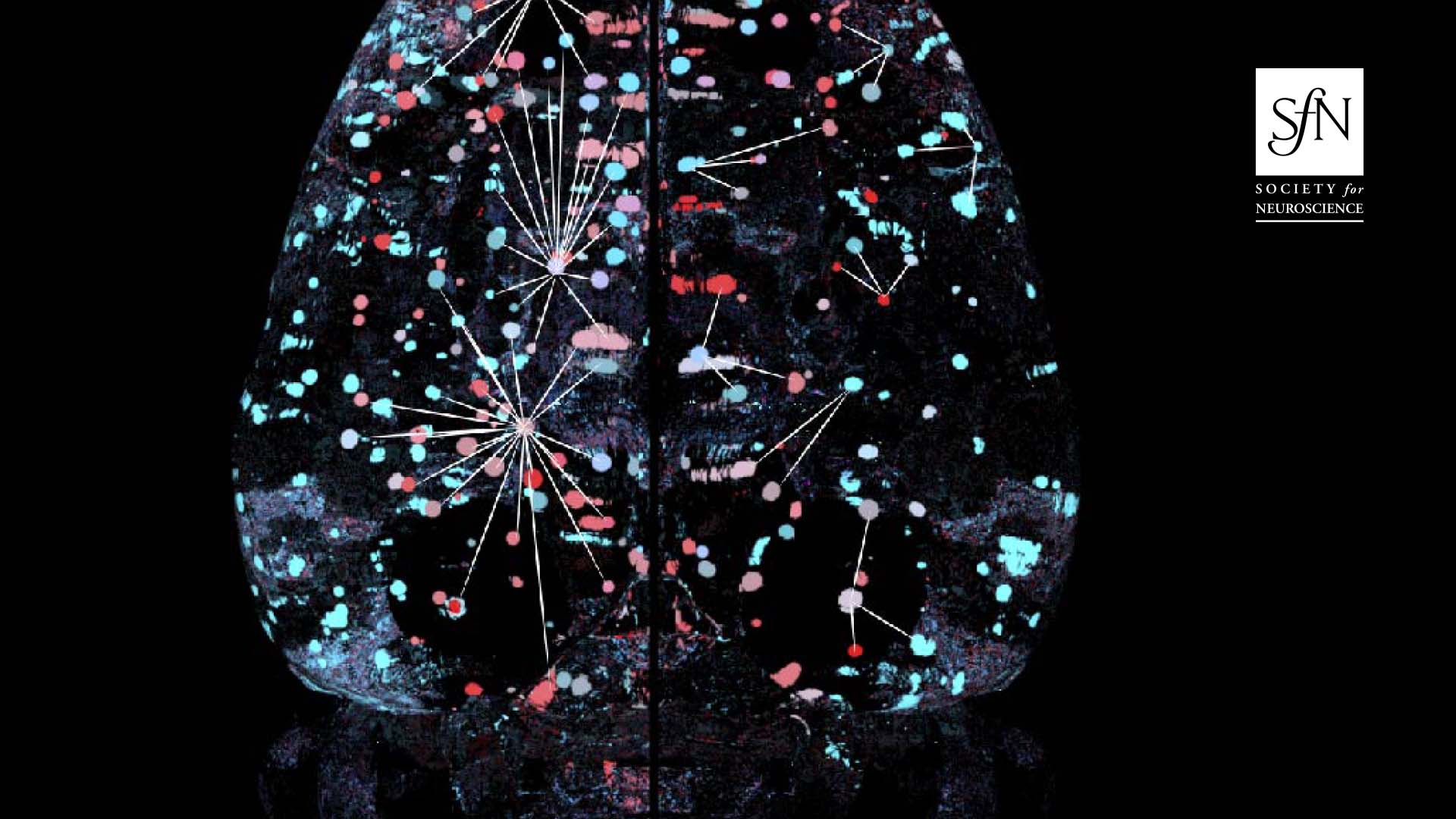
- Characterization of the Brain Functional Architecture of Psychostimulant Withdrawal Using Single-cell Whole Brain Imaging/a>
- Authors: Adam Kimbrough, Marsida Kallupi, Lauren C. Smith, Sierra Simpson, Andres Collazo and Olivier George
- Published: Sept 27, 2021
- Download
-
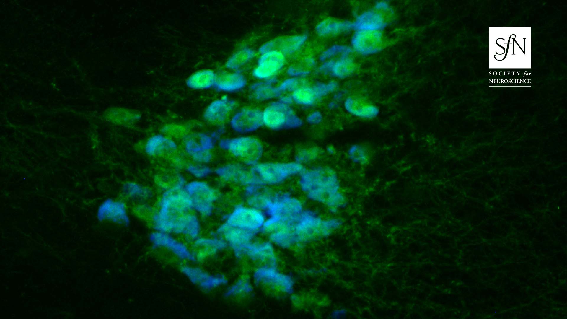
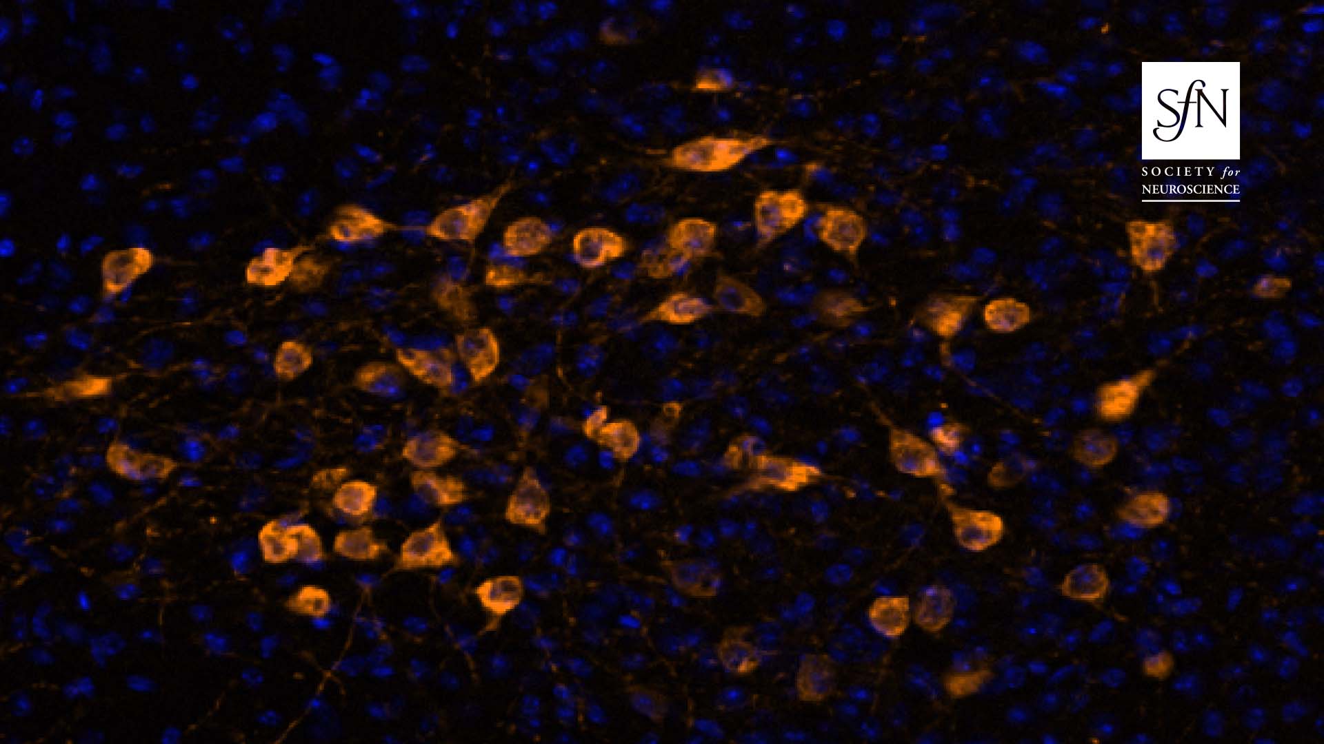
- TRPM4 contributes to subthreshold membrane potential oscillations in multiple mouse pacemaker neurons
- Authors: Keyong Li, Yingtang Shi, Elizabeth C. Gonye and Douglas A. Bayliss
- Published: Nov 3, 2021
- Download
-
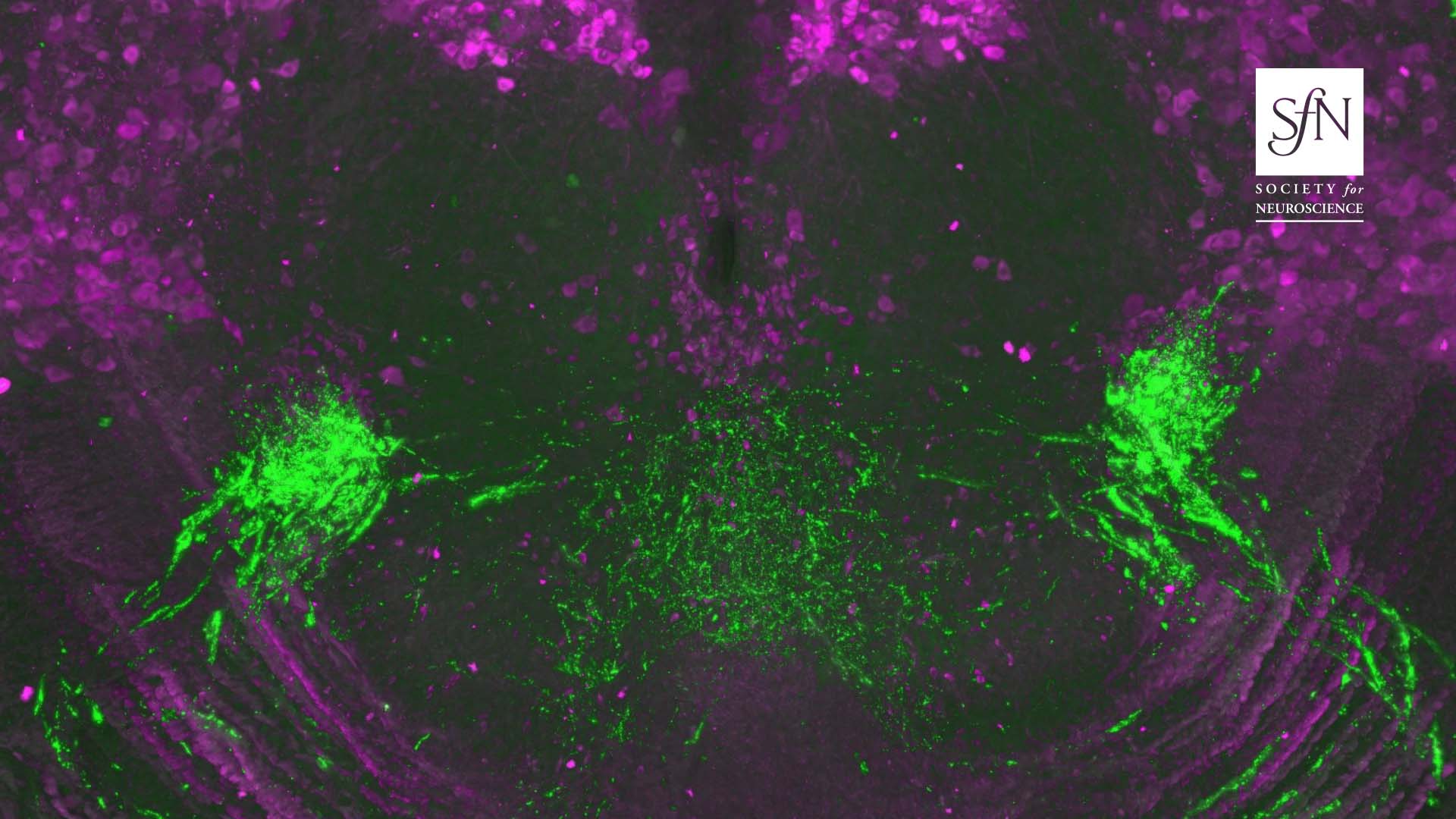
- Off-Target Expression of Cre-Dependent Adeno-Associated Viruses in Wild-Type C57BL/6J Mice
- Authors: Justin J. Botterill, Abdessattar Khlaifia, Brandon J. Walters, Mark A. Brimble, Helen E. Scharfman and Maithe Arruda-Carvalho
- Published: Nov 16, 2021
- Download
-
- Regional Targeting of Bladder and Urethra Afferents in the Lumbosacral Spinal Cord of Male and Female Rats: A Multiscale Analysis
- Authors: J. P. Fuller-Jackson, P. B. Osborne and J. R. Keast
- Published: Nov 12, 2021
- Download
-
- RapID Cell Counter: Semi-automated and Mid-throughput Estimation of Cell Density Within Diverse Cortical Layers
- Authors: Aarthi Sekar, Thiago M. Sanches, Keiko Hino, Matangi Kumar, Juliann Wang, Elisa Ha, Blythe Durbin-Johnson, Sergi Simó and Megan Y. Dennis
- Published: Nov. 1, 2021
- Download
-
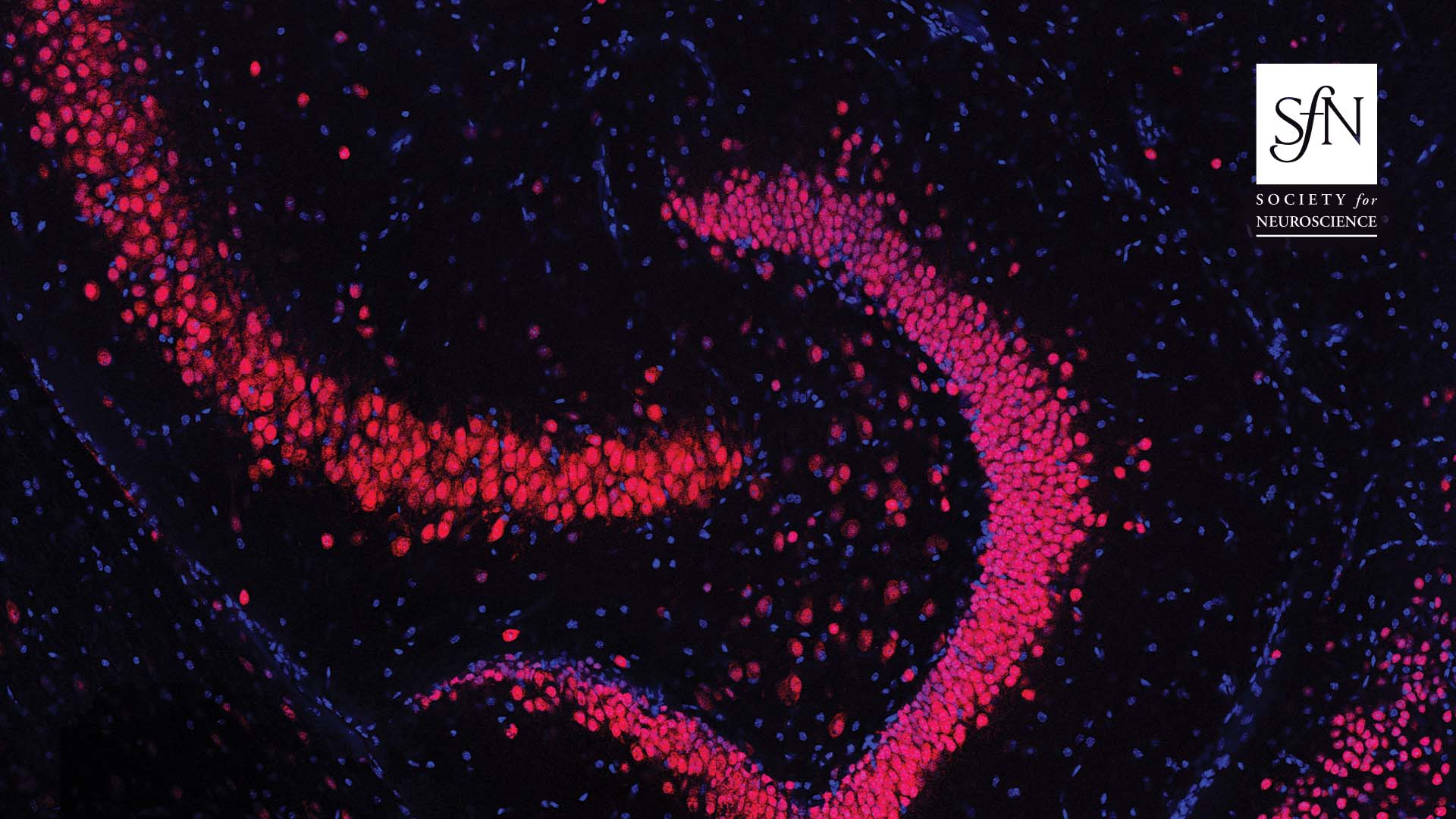
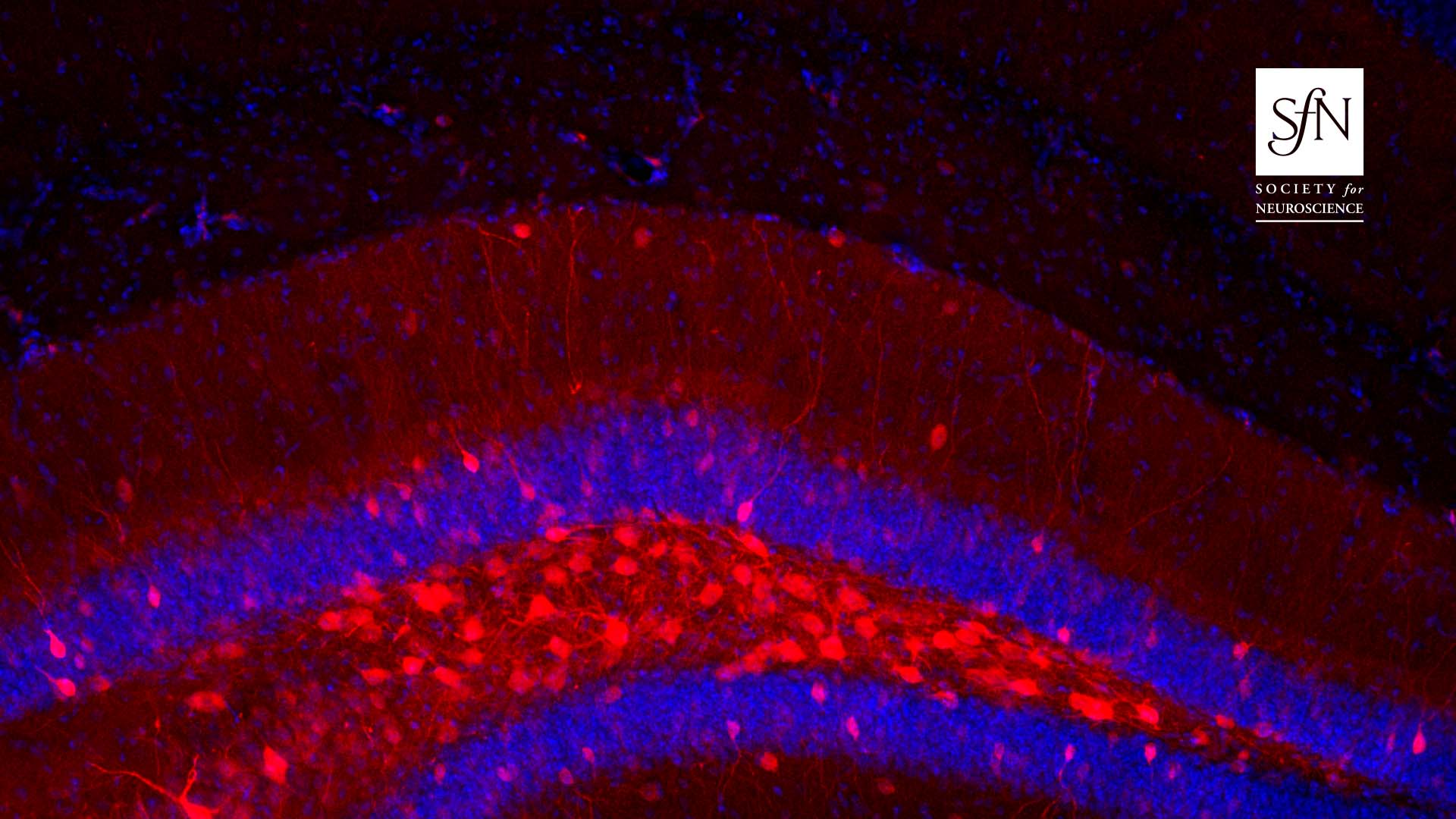
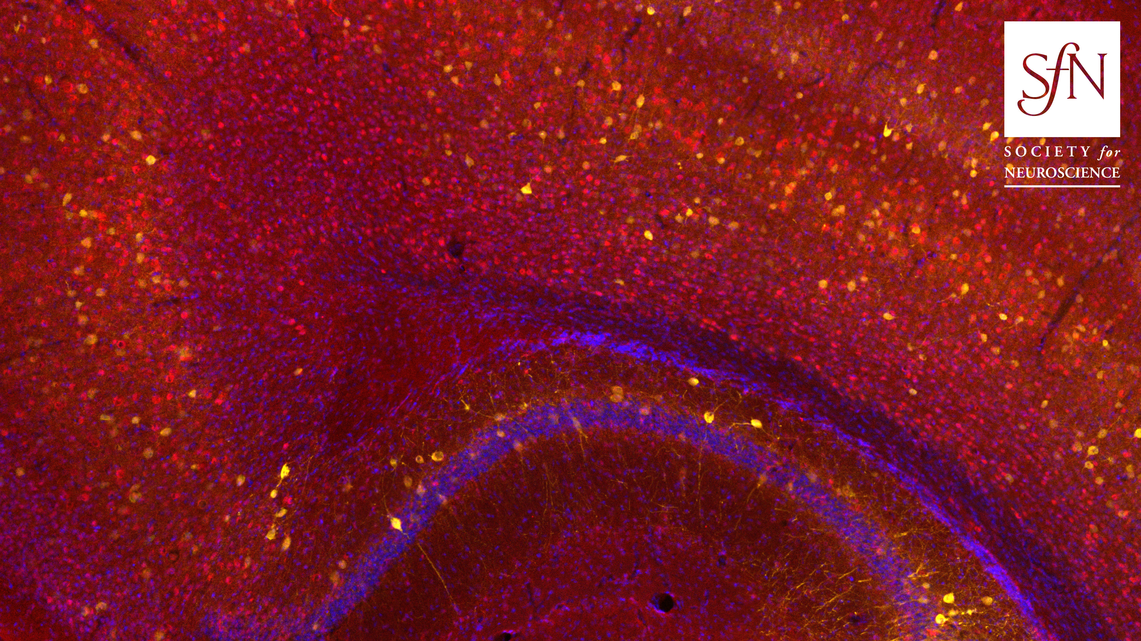
- Slice of Mouse Hippocampus with a Focus on the Dentate Gyrus
- Photo Credit: Jesse Pfammatter
- Authors: Antoine Madar, Jesse Pfammatter, Jessica Bordenave, Erin Plumley, Swetha Ravi, Michael Cowie, Eli Wallace, Bruce Hermann, Rama Maganti and Mathew Jones
- Published: Nov. 17, 2021
- Download
-
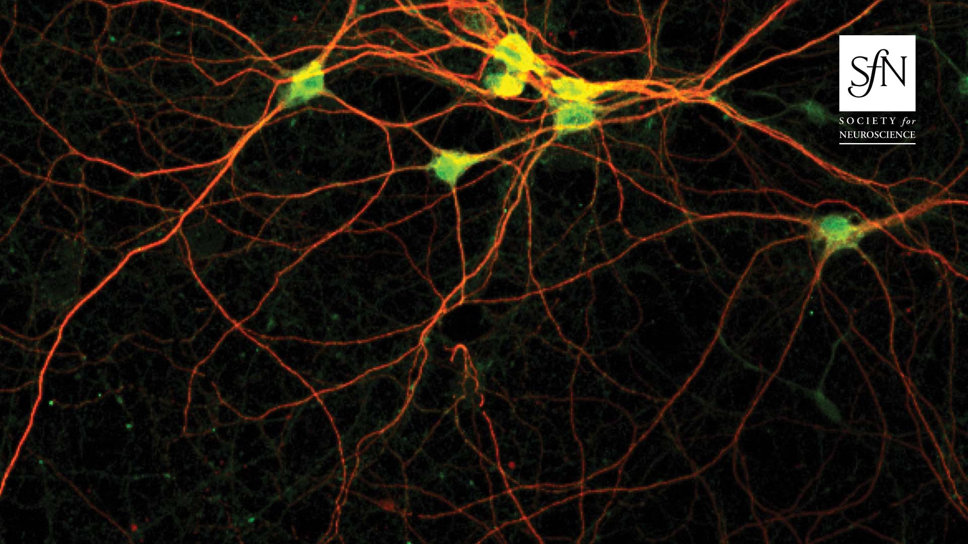
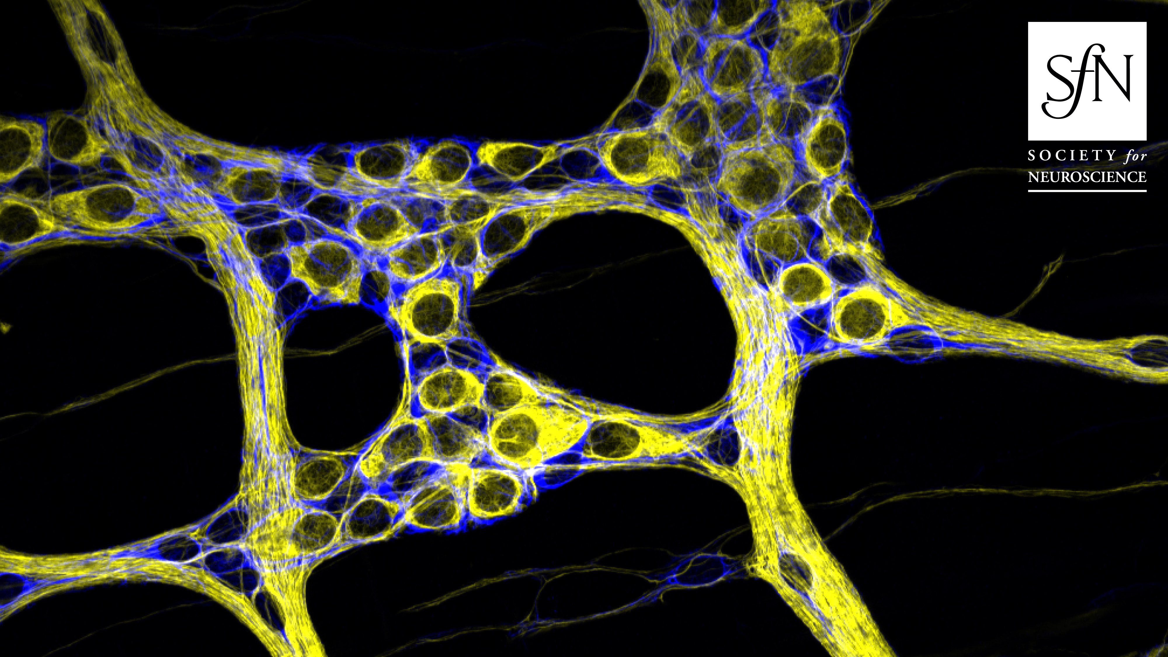
- Cultured Neurons from Mouse Cortex
- Photo Credit: Barbara K. Robens.
- Authors: Susanne Schoch, Anne Quatraccioni, Barbara Robens, Robert Maresch, Karen M.J. van Loo, Silvia Cases-Cunillera, Tony Kelly, Thoralf Opitz, Valeri Borger, Dirk Dietrich, Julika Pitsch, Heinz Beck and Albert Becker
- Published: Sept. 29, 2021
- Download
-
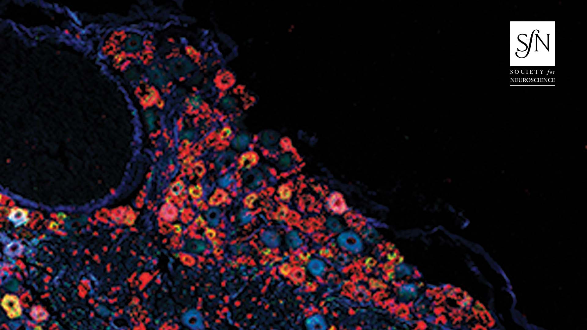
- A 10-μm Thick Section Through a Whole L4 Dorsal Root Ganglion From a Mouse
- Photo Credit: Liam J Peck
- Authors: Liam Peck, Ryan Patel, Paula Diaz, Yolanda Wintle, Anthony Dickenson, Andrew Todd, Margarita Calvo and David Bennett
- Published: Nov. 3, 2021
- Download
-
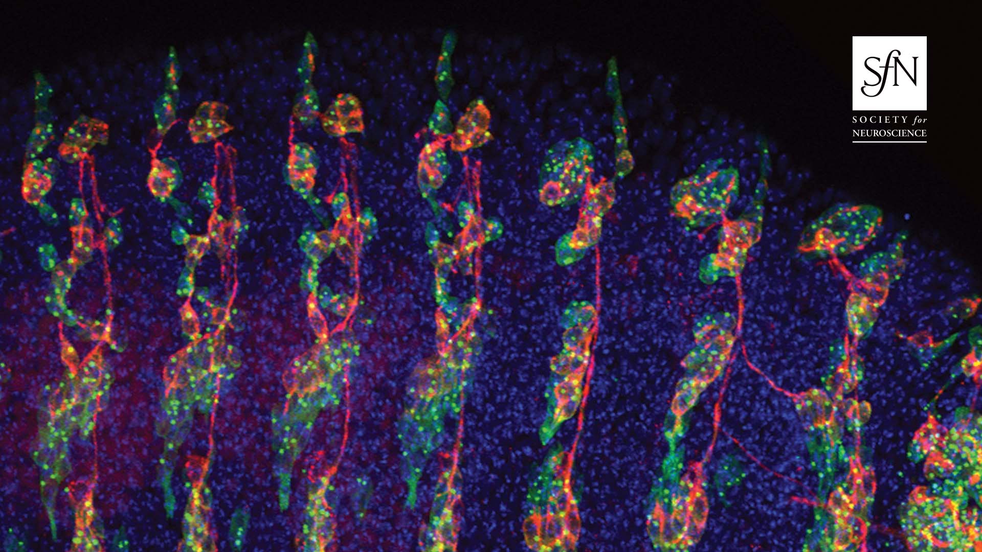
- Expression of miR-263b in a Stage-16 Drosophila Embryo
- Photo Credit: Marleen Klann and edited by Claudio R. Alonso
- Authors: Marleen Klann, A. Raouf Issa, Sofia Pinho and Claudio Alonso
- Published: Oct. 6, 2021
- Download
-
- Cortical Parvalbumin-Positive Interneuron Development and Function Are Altered in the APC Conditional Knockout Mouse Model of Infantile and Epileptic Spasms Syndrome
- Photo Credit: Rachael Ryner and Mary Sommer
- Authors: Rachael F. Ryner, Isabel D. Derera, Moritz Armbruster, Anar Kansara, Mary E. Sommer, Antonella Pirone, Farzad Noubary, Michele Jacob and Chris G. Dulla
- Published: February 22, 2023
- Download
-
- Functional Intraregional and Interregional Heterogeneity between Myenteric Glial Cells of the Colon and Duodenum in Mice
- Photo Credit: Luisa Seguella
- Authors: Luisa Seguella, Jonathon L. McClain, Giuseppe Esposito and Brian D. Gulbransen
- Published: November 16, 2022
- Download
-
- Quantitative Fluorescence Analysis Reveals Dendrite-Specific Thalamocortical Plasticity in L5 Pyramidal Neurons during Learning
- Photo Credit: Ajit Ray
- Authors: Ajit Ray, Joseph A. Christian, Matthew B. Mosso, Eunsol Park, Waja Wegner, Katrin I. Willig and Alison L. Barth
- Published: January 25, 2023
- Download