Images From Neuroscience
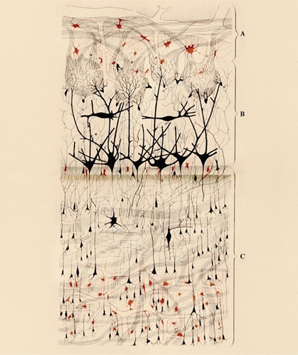
Hand drawn image by Camillo Golgi of a dog's olfactory bulb. Published in: Golgi, C. (1875). Sulla Fina Struttura del Bulbi Olfattorii. Reggio-Emilia: Printer Stefano Calderini.
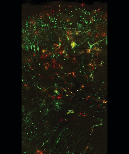
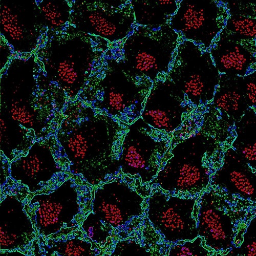
Olfactory bulb of adult mouse brain cells. Mitral cells and interneurons are labeled in green. Groups of glial cells are labeled in red. (2017) Courtesy, with permission: Rebeca Sánchez-González and Laura López-Mascaraque, Instituto Cajal-CSIC, Madrid (Spain).
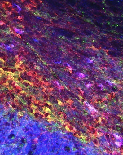
Immunostaining of dopaminergic neurons, which are known to be at most risk of death in Parkinson's disease. The image was processed to provide a watercolor effect. Courtesy, with permission: Murase et al., 2006, JNeuosci, 26 (38) 9750-9760.
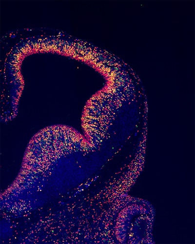
A mouse embryo forebrain on day 11 of development. Nuclei labeled green are daughter cells produced by cell division of neural progenitor cells. Other nuclei are labeled red; double-labeled nuclei are yellow, Courtesy, with permission: Fujimura et al., 2016, JNeurosci, 36 (42) 10908-10919.
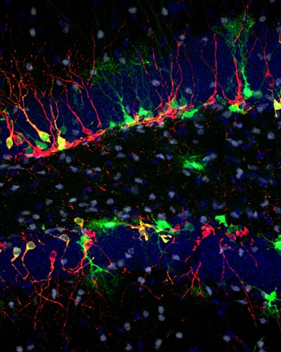
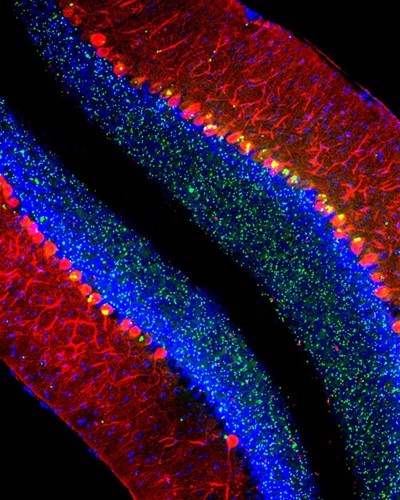
An adult mouse hippocampus with neurogenesis markers. Radial glia-like neural stem cells and their progenies are labeled green. Adult-born neurons and neural stem cells/neural progenitors are stained red and white, respectively. Nuclei are labeled blue. Courtesy, with permission: Kuhn et al., 2018, JNeurosci, 38 (49) 10401-10410.
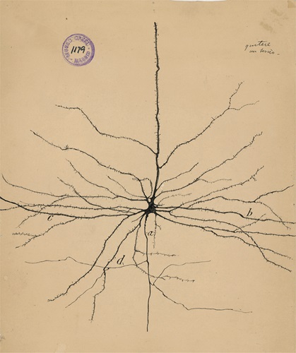
Hand drawn image by Santiago Ramón y Cajal of a pyramidal neuron of the cerebral cortex. (1913) Courtesy of the Cajal Institute, “Cajal Legacy,” Spanish National Research Council (CSIC), Madrid, Spain.
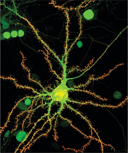
Hippocampal neurons (green) cultured from a mouse model overexpressing Neuroligin-1 (red). Modified from Chanda et al., 2017, JNeurosci, 37 (29) 6816-6836. Courtesy of Soham Chanda.
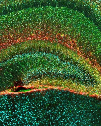
Immunostaining of neural stem cells having a unique morphology called "secondary radial scaffold" (labeled green and red) in the dentate gyrus of a juvenile mouse. Nuclei of neutral stem cells are labeled cyan. (2006) Courtesy, with permission: Noguchi et al., 2016, JNeurosci, 36 (22) 6050-6068.
Cerebellum of a mouse modeling the lysosomal disease late-infantile neuronal cerold lipofuscinosis (CLN2 disease). Purkinje cells are labeled red nuclei blue, and abnormal clumps of the protein p62/Sqtsm1 are green. (2018) Courtesy, with permission: Micsenyi et al., 2013, JNeurosci, 33 (26) 10815-10827.
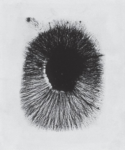
Experiment by Rita Levi-Montalcini observing the effects on a chick embryo 24 hours after exposure to nerve growth factor. (1952) With permission: Levi-Montalcini, 1964, Science, 143 (3602) 105-110.
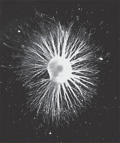
A dorsal root ganglion neuron 24 hours after exposure to nerve growth factor. (2003) Courtesy, with permission: Mills et al., 2003, JNeurosci, 23 (5) 1638-1648.
Ependymal cell basal bodies (red) are clustered in a patch just below the surface of ependymal cells outlined in green. Courtesy, with permission: Mirzadeh et al., 2010, JNeurosci, 30 (7) 2600-2610.