Exploring How Sensory Experience Shapes the Developing Brain
The human brain is immensely complex, comprising billions of neurons and trillions of neuronal connections. Neuronal behavior produces our thoughts, feelings, and actions, and yet how neuronal activity and organization determine our experience of the world has been a challenging question to answer.

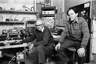
Experiments in the 1960s and 1970s first began to illuminate how neuronal activity and organization produce our complex visual perception of the world. A pioneering study in 1959 launched the Nobel Prize–winning collaboration between David Hubel and Torsten Wiesel, who used the then-new electrophysiological technique of recording the activity of single neurons to reveal the pattern of organization of cells that process vision and how this organization gives rise to visual perception. Both of them later served as presidents of SfN.
Hubel and Wiesel demonstrated that visual experience early in life has long-lasting effects on the structure and function of the brain’s visual processing system. They discovered that when an animal lacked vision in one eye during a critical developmental period, neurons in the primary visual cortex expanded into the regions that normally would have received input from that eye. This showed that the brain’s cortex changes during development in response to experience and highlighted the importance of sensory input during critical periods of development.
From their studies, Hubel and Wiesel inferred that the structure and function of brain connections is highly dynamic during development. At the time, however, there was no way of visualizing those dynamics in real time. This made it difficult to decipher the molecular and cellular mechanisms underlying experience-dependent changes in the brain.
Hollis Cline, SfN past president and professor at the Scripps Research Institute, has unlocked some of the secrets of how experience changes connections in the brain. Cline studies the development of the visual system in Xenopus tadpoles. One advantage of studying these animals is that they are transparent in the early stages of their lives, allowing researchers to peer into their brains to see individual neurons.
Using this model system combined with technological advances such as in vivo imaging with high-resolution microscopy, Cline has demonstrated a range of effects of visual experience on the development and plasticity of the visual system. For instance, Cline's group showed that brain connections grow and withdraw in a matter of hours and days during development. Projections from neurons called dendrites allow neurons to send signals to other cells. Growing dendrites produce many branches, and as new branches grow, they form connections, called synapses, to other neurons.
"We saw that there is a constant dynamic, with branches in these complex trees adding and retracting at a rapid rate," Cline said. "A tiny fraction of those newly added branches are stabilized and persist. Prior to taking these time-lapse images, we would never have guessed this would be the process by which a cell would grow and develop these extremely elaborate dendritic branches.”

Visual experience early in life has long-lasting effects on the structure and function of the brain’s visual processing system.
Cline’s research revealed that both new dendrites and new synapses are transient, with some growing stronger but many more than expected withering away. She has also contributed data that show that the general principles of brain development demonstrated in vertebrate animal models hold true in humans.
Experiments such as Cline’s have been essential for testing ideas about how the developing brain builds and maintains new connections as an animal is exposed to different sensory information. Research like this also has a lot to tell us about what happens when something goes wrong during brain development.
“Brain circuit formation is absolutely fundamental to animals and people being able to grow and learn and carry out motor tasks,” Cline said. “If the brain doesn’t grow, then none of these things happen.”
A tragic example of disrupted brain circuit formation is the recent epidemic of Zika virus, which infects and kills brain cells in fetuses, causing microcephaly. Understanding how the brain grows could have implications for Zika virus as well as for neurodevelopmental diseases such as autism spectrum disorders.
While unique experimental models and technological advances in imaging and manipulating the brain are yielding insight about which Hubel and Wiesel could only speculate, many neurodevelopmental diseases are still not well understood. Scientists like Cline hope that unraveling the core deficits of these diseases at a cellular and molecular level will lead to better treatment options.
“The kinds of research we have done and the contributions that we have made to brain science would have been impossible without an experimental model like Xenopus tadpoles,” Cline said, “but unless we as scientists continue using a range of experimental systems that allow us to address questions of interest, we’re going to slow down our own progress.”


